Biopsy of a mass in our veterinary patients is one of the most powerful tools in veterinary oncology because it turns a ‘lump’ into a specific diagnosis with real prognostic information that can guide treatment decisions and expected outcomes for those clinicians dealing face-to-face with their patients and clients.
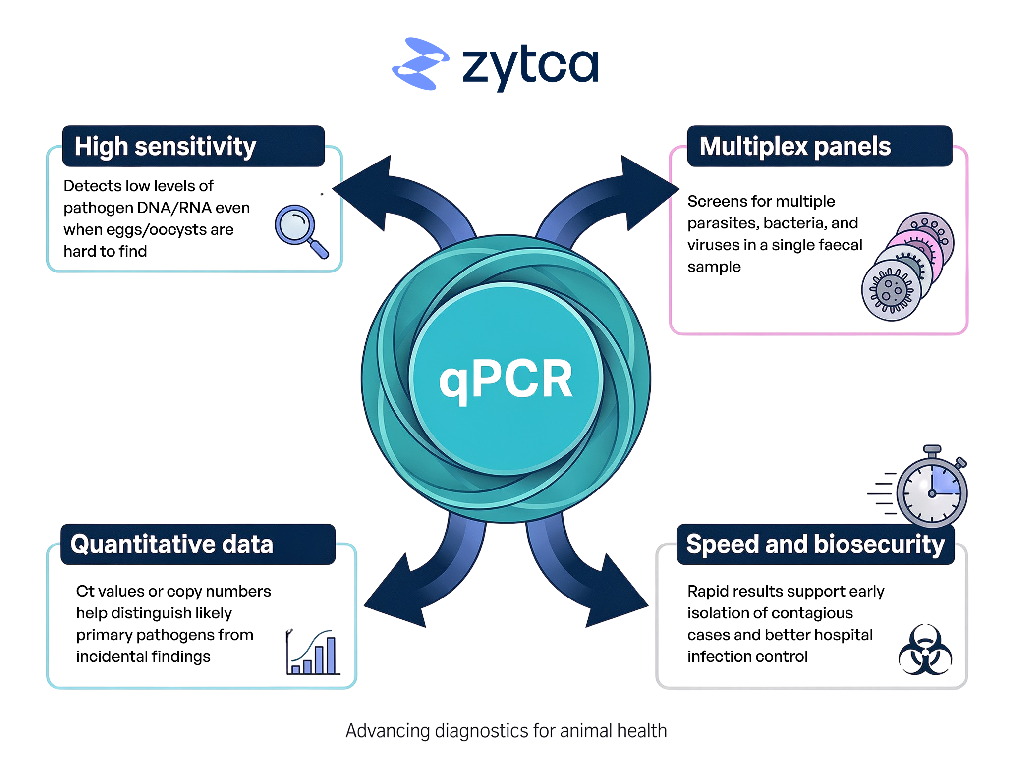
Meuten et al (2021) published ‘The International Guidelines for Veterinary Tumor Pathology: A Call to Action’ in Veterinary Pathology, which sets out standardised methods for assessing tumours histologically in animals. Consistent biopsy handling, staining and reporting (including mitotic count, margins, necrosis and invasion) are essential if clinicians are to rely on tumour grade and other histologic feature to predict survival, recurrence and metastatic risk. This paper highlights microscopic features for common examples such as canine soft tissue sarcomas and melanocytic tumours, amongst others. Ultimately, the overlying message through the paper is that without high-quality, standardised histopathology of biopsies, veterinary oncology cannot generate robust evidence to improve patient care. At Zytca Animal Health we are developing AI-assisted histopathology reporting by board-certified pathologists which allows our reports to utilise digital images of submitted biopsies and report even more accurate features of neoplastic and non-neoplastic tissues.
💡Accurate tumour type and grade
Biopsies are able to differentiate between benign and malignant lesions and often identified the exact tumour type, which is often impossible with imaging or cytology alone. In attributing type and grade to the lesion, this strongly influences the expected behaviour, from slow-growing, locally confined masses to highly metastatic cancers, and therefore determine how aggressive surgery and/or adjunctive therapy should be.
💡Surgical planning and margins
This paper highlights the strong importance of margin assessment from histologic examination of excised tumours is as often as important as the exact diagnosis itself. This is because incomplete margins (where the lesion extends to the cut edge of the tissue) are strongly associated with local recurrence. Knowing in advance (from an incisional (portion of the lesion) biopsy), whether a mass is likely to need wide margins, limb-sparing, or referral-level surgery can prevent under- or over-treatment.
💡Prognosis and follow-up
Microscopic features of a mass, such as mitotic count, necrosis and lymphovascular invasion allows pathologists to place an individual patient into published prognostic categories (e.g., high or low grade mast cell tumours, or melanocytic tumours with defined mitotic cut-offs linked to survival). This helps clinicians provide realistic survival estimates, decide on staging tests and plan follow-up intervals and monitoring strategies.
Ultimately, appropriate tissue sampling of lesions allows clinicians to avoid both overtreatment or benign or low-grade tumours and undertreatment of more biologically aggressive neoplasms. Over time, larger, well annotated datasets linking standardised histology with clinical outcome are only possible when tumours are biopsied and assessed in a consistent way. Utilising an AI-assisted platform such as AniPathTM, allows critical features of grading schemes to be more accurately and consistently applied. This in turn refines prognostic models and treatment protocols over time. In considering the individual patient, this means that a relatively simple procedure (incisional or excisional biopsy) can transform an uncertain situation into an evidence-based plan that incorporates appropriate surgical margins, considered use of chemotherapy or radiotherapy and tailored follow-up based on validated histologic risk factors.
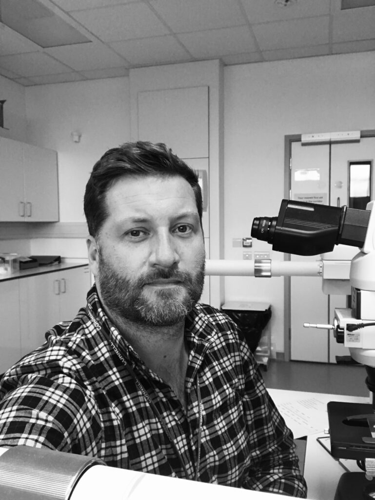
Dr Marvin J. Firth is the Director of Veterinary Pathology at Zytca Animal Health. He is a RCVS and American specialist in Anatomic Pathology and has an interest in neoplastic and infectious disease, digital pathology and artificial intelligence and communication in science.
Reference:
Meuten DJ, Moore FM, Donovan TA, et al. International Guidelines for Veterinary Tumor Pathology: A Call to Action. Veterinary Pathology. 2021;58(5):766-794. doi:10.1177/03009858211013712